Open a medical journal, a news website, or a radiology blog, and surely artificial intelligence (AI) will be mentioned. AI is making waves in medical imaging. This can be exciting, as AI offers many opportunities to improve the radiology workflow; however, it also poses a challenge. How do you start working with AI in clinical practice without having to deal with a tiring amount of implementation hassle?
Below are our top 8 learnings to help with a smooth implementation of AI solutions at your department.
Tip 1: Include non-imaging clinicians in the radiology AI implementation process
 If the radiology department is changing the way medical imaging results are delivered or if extra information is included in radiology reports, referring physicians will want to be informed. Desisting from doing so may cause unnecessary friction in the implementation process.
For example, Quantib® ND Plus, our AI radiology software for measuring brain atrophy, will provide neurologists with objective volume measurements instead of the visual scoring (neuro)radiologists currently deploy. If your neurologists are not informed before implementation, this change can cause confusion. Involving the neurologists in the implementation process will prevent a wave of questions and they will most likely be enthusiastic about the improvements proposed by the radiology department.
Ideally, all stakeholders are on board even before opening discussions with AI companies. The sooner everyone is on the same page, the less obstacles are expected during purchasing and implementation.
Tip 2: Tell your patients you are using radiology AI software and why
 Involving all stakeholders in the AI implementation process goes for the patients as well. Once the AI solution is up and running, it will (it should!) definitely affect your patients. Why not share information about the improvements the radiology department implemented and how these will benefit them? Experience tells us that using AI to enhance your diagnostic process can lead to a significant increase in patient volumes, because they value the additional detailed information AI results can offer.
How to inform your patients? Publish a press release about the new AI software and its benefits for patients and healthcare professionals. Sharing this on social media will bring positive attention to your facility. Organize an informational session for patients, relatives, and/or caretakers. Do not hesitate to collaborate with your AI vendor for this, because, in most cases, they will be happy to assist.
Tip 3: Do not be scared to delegate the radiology AI implementation!
 Many imaging departments have radiographers, physician assistants, or nurses who are willing to stick out their necks and learn new things. Turn first steps into the world of AI into a win-win situation for the whole department by having these people perform (part of) the AI analyses. Radiologists gain time to focus on specialized tasks and radiographers enrich their routine work with more responsible tasks which they might not be able to perform in competing facilities. The deal works for everyone.
Tip 4: Do not underestimate the value of service in radiology AI
 Getting started with radiology AI will take time. Yes, there may be technical issues, but in our experience, most time consuming issues are due to people needing to adjust to new ways of working.
This requires an AI vendor that is willing to invest in the relationship and assist with making this transition period as smooth as possible. So be picky when selecting the right vendor and choose a company with a well-oiled service team, short lines of communication, and a dedicated customer contact employee.
Tip 5 : Check for suitable radiology AI reimbursement codes
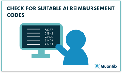 Purchasing the newest radiology AI solutions for your imaging facility can be an uphill battle with your financial department. To simplify your business case, check whether any suitable reimbursement codes are available that could also apply for AI analyses. This could make the software pay for itself. For example, in the USA, CPT code 76377 reimburses for various exams applying image post processing on an independent workstation. It might just make your discussion with the financial department a lot easier.
In Europe, this is a bit more tricky. As far as we know, there is no similar arrangement available. However, other elements can enhance your business case. Does the software provide easy to understand reports such that these are straightforward for referring physicians to understand? Perhaps, in that case, it is not crucial for a radiologist to attend the multi-disciplinary team meetings (MDTs) to present the imaging results. Hence, the radiologist is able to save a lot of time, leading to financial savings (or an optional increase in imaging volume). Some of our Quantib® ND customers use the software and corresponding reports in this way.
Tip 6 : Consider the radiology AI company’s pipeline
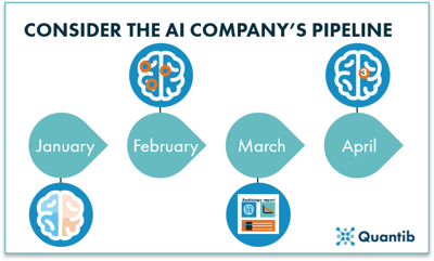 Think beyond your current purchase and investigate the product pipeline of different radiology AI vendors. During the purchasing process, you will most likely compare similar products from different companies. It is worthwhile to discuss with each vendor what their planned updates look like and for which period they are scheduled. The AI software of vendor A might be best right now, but if the package of vendor B will surpass that level in 2 months, you are better off buying the latter.
Tip 7 : Radiology AI software results versus current clinical practice
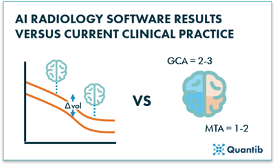 One of the most important things to keep in mind when you are implementing a radiology AI solution is that the results of the algorithm, in most cases, cannot be compared in a straightforward manner with a visual assessment. Algorithms are capable of detecting small changes not yet visible to the human eye, or comparing those results to databases of thousands of healthy subjects, thus bringing a whole new perspective into your reporting room.
For instance, Quantib® ND, our neurodegeneration software, provides brain structure volumetry. Current clinical practice includes atrophy assessment by using visual scoring systems, such as the global cortical atrophy (GCA) scale, the medial temporal lobe atrophy (MTA) scale, or the Koedam score for parietal atrophy. These systems are based on a visual assessment of grey matter atrophy, ventricle size, gyri, sulci, etc. Of course this is not the same as having quantified volumes and knowing what a certain volume (decrease) means. This does not mean the algorithm is unreliable, but that it provides a slightly different type of output compared to what radiologists have been used to working with so far.
Tip 8 : Do not be shy. Give feedback on the radiology AI solutions!
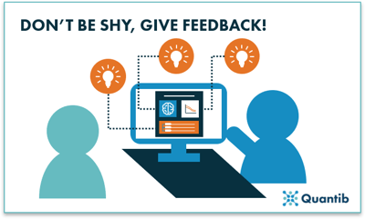 Early adopters will shape the future of radiology. Radiologists that implement artificial intelligence today can proactively provide feedback and, therefore, contribute significantly to decisions that are made today on how radiology AI will be designed and implemented in the coming years. AI companies exist of smart and motivated people, working together to improve patient outcomes. However, vast clinical experience is not always part of the permanent team. Therefore, direct feedback from the end-user is always highly appreciated. Feedback users may deem trivial can be the start of an amazing improvement coming to your clinic in a new update!
Are 8 tips for radiology AI implementation not enough?
Every implementation process is different. Some of the tips mentioned above may prove useful when you are getting your own radiology AI software up and running, and some may not apply to your situation. Did we forget to cover a question you have? Or are you curious to learn more about how we implement our software solutions? Don’t hesitate to reach out to us!
